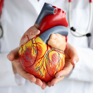
Our pulmonology team deals with everything related to the functioning of the respiratory system: lungs, bronchi, pleura and trachea.
CHRONIC OBSTRUCTIVE PULMONARY DISEASE
Chronic obstructive pulmonary disease (COPD) is a progressive, non-infectious lung disease that affects the respiratory system and makes breathing difficult. COPD is also known as chronic obstructive pulmonary disease (COPD).
COPD includes chronic bronchitis, emphysema and other similar lung diseases.
Chronic bronchitis is a disease of the airways in the lungs. It is characterised by coughing, mucus production and chest congestion. Chronic bronchitis is more common in smokers than in non-smokers.
Chronic bronchitis is often associated with asthma or emphysema. The combination of chronic bronchitis and emphysema is called chronic obstructive pulmonary disease (COPD).
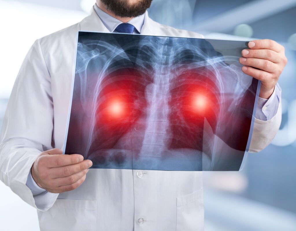

ASTHMA AND AIRWAY ALLERGIES
Asthma and respiratory allergies (take in charge in our Pulmonology department) are defined as irregular narrowing of the airways caused by inflammation of the bronchial tubes.
This can be triggered by various factors, such as environmental irritants (pollution, dust/smoke) and cigarette smoke, infections or physical exercise. Asthma and respiratory allergies affect about 300 million people worldwide, especially young women with seasonal allergies.
Asthma is a chronic (long-term) disorder of the airways. Asthma can range from mild to fatal and affects people of all ages. It causes inflammation, narrowing of the airways and an increased risk of bronchospasm (constriction).
RESPIRATORY INFECTIONS
Our respiratory department supports all respiratory infections, which are infections of the upper respiratory tract, including the nose, throat and sinuses, caused by bacteria and viruses.
Respiratory infections can cause symptoms such as fever, headache, coughing and sneezing. Respiratory infections are caused by a wide variety of pathogens with different infection mechanisms, causing different types of disease.
The most common causes of respiratory infections in adults are:
- The common cold (virus)
- Influenza (virus) – flu season occurs during the winter months; the most severe symptoms occur within two days of exposure to the virus.
- Bronchitis (virus or bacteria) – inflammation of the walls of the large airways; usually caused by a virus that attacks the walls of the bronchi; symptoms include cough with phlegm and shortness of breath.
- Pneumonia (bacteria) – inflammation of the lung tissue that can lead to fluid accumulation in the lungs; usually caused by bacteria, but can also be caused by viruses or fungi.
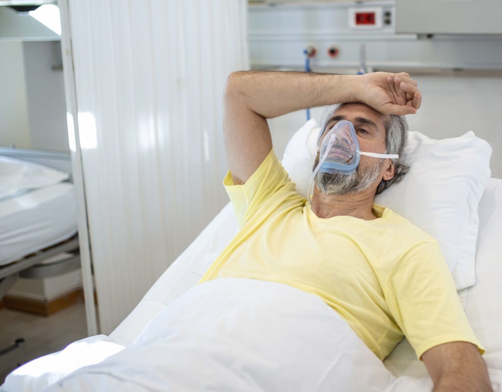
BRONCHOPULMONARY TUMOURS
A bronchopulmonary tumour (take in charge in our Pulmonology department) is a type of lung neoplasm (the formation of abnormal tissue in the respiratory system) that comes from the cells that line the airways.
There are two types of bronchopulmonary tumour, benign and malignant. Benign tumours do not spread to other parts of the body and can be treated with medical or surgical intervention, while malignant tumours have the potential to spread if left untreated.
Bronchopulmonary tumours include:
- Lung cancer – cancer that develops in lung cells.
- Tracheobronchial carcinoid (TBC) – A tumour that develops in the bronchi and has similar characteristics to neuroendocrine tumours (NETs).
- Bronchioloalveolar carcinoma (BAC) – Cancer that develops in the cells that line the airways. It may also be called adenocarcinoma, lymphoma or squamous cell carcinoma.
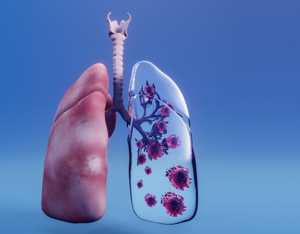

RESPIRATORY FAILURE
Respiratory failure (take in charge in our Pulmonology department) is a life-threatening condition in which the lungs cannot supply enough oxygen to the body.
Respiratory failure can be acute (rapid onset) or chronic (slow progression). Causes include pneumonia, influenza, cancer, heart failure and certain lung diseases such as emphysema and chronic obstructive pulmonary disease (COPD).
Emphysema is a chronic and progressive lung disease that causes shortness of breath. It causes the walls of the alveoli (air sacs) to break down, reducing the body’s ability to exchange oxygen and carbon dioxide, leading to a feeling of tiredness and shortness of breath.
SLEEP APNOEA
Our pulmonology department supports sleep apnoea, a sleep disorder in which breathing stops and starts repeatedly during sleep.
The most common cause is a drooping of the soft tissues in your airways, which blocks them and interrupts your breathing.
Sleep apnoea usually has no symptoms during the day, but it can lead to problems such as high blood pressure, depression and heart disease.

TUBERCULOSIS
Tuberculosis (TB) is a disease that can affect the lungs and other parts of the body. It usually affects the lungs, but can also affect other parts of the body, such as the kidneys, spine and brain. It is caused by a germ called Mycobacterium tuberculosis.
Tuberculosis (take in charge in our Pulmonology department) is spread through tiny droplets in the air when an infected person coughs or sneezes. It can also be spread by sharing objects such as drinking glasses or kitchen utensils with someone who has tuberculosis.
The signs and symptoms of TB are:
– A bad cough that lasts for 3 weeks or more
– Painful breathing or chest pain
– Coughing up blood (haemoptysis)
– Loss of appetite and weight loss
PLEURISY
Pleurisy is an inflammation of the pleura, the membrane lining the inside of the chest cavity. The pleura* is a thin layer of tissue that covers both lungs. When you breathe, air passes between the lungs and the chest wall.
Pleurisy is a painful condition that causes sharp or throbbing sensations in the chest. You may also feel short of breath or have an increased heart rate.
Pleurisy can be caused by an underlying disease or injury. It can also occur as a complication of another condition, such as pneumonia or heart failure.
If you have mild pleurisy, your doctor may recommend painkillers and nursing care. Severe cases are treated with anti-inflammatory drugs, antibiotics and, if necessary, surgery.
EMPHYSEMA
Emphysema is a chronic lung disease that causes shortness of breath, wheezing and other symptoms.
Emphysema is an abnormal enlargement of the air sacs in one or both lungs. Lung tissue can be damaged, which can cause permanent damage to the lungs.
As emphysema progresses, you may experience shortness of breath during daily activities such as climbing stairs or getting dressed. Eventually, your breathing will become so difficult that you will need extra oxygen (in the form of a nasal cannula) when you are awake.
SHOULDER PLEURAL
A pleural effusion (take in charge in our Pulmonology department) is an accumulation of fluid in the pleural space, which is the space between the lungs and the chest wall. The pleural space contains two thin membranes that produce a lubricating liquid called pleural fluid. Excess fluid can interfere with lung function and cause shortness of breath, cough, chest pain, fever and weight loss.
Pleural effusions can be caused by heart failure or cancer, but most are caused by infections such as pneumonia or tuberculosis.
The main treatment for pleural effusion is to drain it with a needle or chest tube. In some cases, an antibiotic may be given to treat the underlying infection.
PNEUMOTHORAX
A pneumothorax is a build-up of air in the space between the lungs and the chest. It is most often caused by an injury to the lungs, such as a broken rib or a deep cut in the chest.
A pneumothorax can also be caused by a perforation of the lung (pneumothorax) or a spontaneous rupture of a lung (hemothorax).
The air in the chest cavity prevents the lungs from expanding and breathing properly, which reduces the amount of oxygen in the blood. Pneumothorax can be fatal if not treated properly.
SARCOIDOSIS
Sarcoidosis (take in charge in our Pulmonology department) is a disease in which non-cancerous masses called granulomas form in the body’s tissues. Granulomas are areas of tissue that have become inflamed or irritated.
The cause of sarcoidosis is unclear, but it seems to be related to a problem with the immune system. The immune system is the body’s defence against foreign invaders such as bacteria, viruses and fungi. It also helps to protect against cancer cells.
Sarcoidosis can affect any part of the body, but it most commonly affects the lungs, eyes, skin and lymph nodes (glands that store lymphocytes).
The disease usually heals after a few years without treatment. But some people – especially those who have difficulty breathing – may need treatment to help them breathe and feel better.
We present to you some articles relating to pathologies treated in our Mediterranean clinic, with an effective and efficient medical and paramedical team.
Thanks to the latest advances in pulmonology, our breath is coming back to life. Modern pulmonology offers innovative solutions to improve our breathing capacity and general well-being.
Découvrez tout ce que vous devez savoir sur la rhinite allergique, y compris ses symptômes, ses causes et les traitements disponibles, dans ce guide complet.
Lung cancer is a serious illness that can affect people of all ages and backgrounds. In this comprehensive guide, you’ll learn about the symptoms, causes, and treatments of this disease, as well as tips for preventing lung cancer.
Do you suffer from sleep apnoea or think you might have it? Discover the symptoms, causes, and treatments in this comprehensive guide.
TESTS RELATED TO PNEUMOLOGY
Respiratory Functional Testing (RFT) is a method of examining the respiratory system based on the patient’s complaints and answers to questions.
RFT can be used to assess patients with various respiratory problems such as asthma, chronic cough, bronchitis, pneumonia, wheezing and other signs of airway obstruction.
RFT can be performed by healthcare professionals without formal training in respiratory physiology or pathology, including general practitioners and nurses.
This simple approach allows clinicians to quickly identify many conditions that can cause symptoms and determine which test(s) should be performed to confirm the diagnosis.
Why use the Respiratory Functional Test?
Respiratory functional test (RFT) allows healthcare professionals to identify patients who may have a respiratory abnormality without having to perform expensive tests such as a pulmonary function study or fibre-optic bronchoscopy. It also makes it possible to determine whether the patient has multiple pathologies at the root of their symptoms or just one specific problem.
Polygraphy (take in charge in our Pulmonology department) is a medical procedure used in the diagnosis and treatment of lung diseases. It is also known as plethysmography or plethysmograph. Polygraphy is a method of measuring changes in lung volume in response to various stimuli. It can be used to measure lung function and quality of breathing, and to determine whether a patient has asthma or other lung disease.
Polygraphy measures the amount of air exhaled after inhalation. This measurement is called vital capacity. Normal vital capacity varies with age, sex and height, but should be between 3 and 6 litres (or 3 to 6% of body weight). A person with asthma may have a reduced vital capacity. In addition to vital capacity, polygraphy can measure other aspects of breathing such as respiratory rate (number of breaths per minute), current volume (volume of each breath) and respiratory frequency variability (change in respiratory rate from one minute to the next).
Why use the polygraphy?
Polygraphy can help your doctor diagnose some common lung diseases, including asthma, chronic obstructive pulmonary disease (COPD) and lung cancer.
You ask, our teams answer.
F.A.Q
Pulmonology is the study of the lungs. It is a medical speciality that focuses on the diagnosis and treatment of diseases of the respiratory system.
A pulmonologist (from the Greek word pneuma, meaning “air”) is a doctor who specialises in diseases of the respiratory system.
The pulmonologist’s main role is to diagnose and treat people with lung diseases such as asthma, chronic obstructive pulmonary disease (COPD), pneumonia, lung cancer and many other conditions. They can also treat people with heart failure and other cardiovascular diseases that affect breathing, such as emphysema or bronchiectasis.
The field of pulmonology is not the synonym of pulmonology, which focuses on the treatment of diseases and disorders of the respiratory tract. The field of pulmonology includes subspecialities such as intensive care, sleep medicine, pulmonary medicine, thoracic surgery, paediatric pulmonology and others.
If you have a lung disease, Pulmonologists are specialists in the diagnosis and treatment of all types of respiratory diseases. They can help you manage asthma, COPD, lung cancer and other conditions.
If you have chest pain, Pulmonologists can assess your symptoms and determine whether they are caused by heart or lung problems. They can also treat heart failure associated with pulmonary hypertension or other lung diseases. If you have a chronic cough or shortness of breath. Pulmonologists are experts in diagnosing and treating these symptoms, which can be caused by infections such as pneumonia or bronchitis, viral infections such as influenza or tuberculosis, or chronic obstructive pulmonary disease (COPD). They are also used to treat tuberculosis and other lung infections.
If you want to stop smoking, Pulmonologists can help you stop smoking through programs that include advice on healthier lifestyles, nicotine replacement therapy and other medicines.
If you have certain types of cancer, such as lung cancer, which requires specialist care from a pulmonologist with experience of this type of cancer.
Respiratory failure (take in charge in our Pulmonology department) is a severe form of acute respiratory distress syndrome (ARDS). It is a serious, life-threatening condition. It occurs when a person’s lungs do not get enough oxygen.
Respiratory failure can be caused by:
– Damage to the chest cavity (lungs) or lung tissue (lung)
– Obstruction of the airways by foreign bodies or swelling (pneumonia)
– Infection of the lungs (pneumonia)
– Respiratory failure can be acute (sudden onset) or chronic (developing over time). Respiratory failure can also result from underlying lung disease, such as pneumonia or heart failure.
Acute respiratory failure occurs when the lungs cannot exchange oxygen and carbon dioxide efficiently, resulting in hypoxaemia (low levels of oxygen in the blood) and hypercapnia (high levels of carbon dioxide in the blood). Hypoxaemia can cause cyanosis (bluish discolouration of the skin due to lack of oxygen in the blood), shortness of breath, confusion, disorientation, loss of consciousness and even death if not treated promptly with supplemental oxygen or mechanical ventilation. Acute respiratory failure is often caused by pneumonia or other infections of the lungs, or by severe asthma attacks.
Chronic respiratory failure occurs when a person’s lungs don’t work properly for a long time due to long-term damage caused by conditions such as emphysema or chronic obstructive pulmonary disease (COPD). Chronic respiratory failure often develops slowly over time, but can become suddenly acute when it progresses rapidly or manifests as seizures.
Legionellosis is a disease caused by the bacterium Legionella pneumophila. The disease can be transmitted by inhaling contaminated aerosols from water sources such as air conditioners, showers and fountains.
Symptoms of legionellosis may include:
- Fever
- Chills
- Muscle aches
- Headaches
- Tiredness
- Chest pain (may be severe)
Bronchitis (take in charge in our Pulmonology department) is a lung disease that causes inflammation of the bronchial tubes, the airways leading to the lungs. The main symptoms of bronchitis are coughing and wheezing. The cough may be dry or produce mucus.
The severity of bronchitis can range from a mild cough lasting a week or two to a severe, long-lasting condition. Acute bronchitis often starts with an infection of the upper airways (nose and throat), followed by swelling and irritation of the lower airways (bronchi). These irritated airways then produce more mucus than usual, which can make it difficult to breathe deeply without coughing.
Chronic bronchitis, which lasts three months or longer, occurs when there is a long-term change in lung function. This happens when the lining of the airways is damaged by previous infections or by exposure to irritants such as tobacco smoke, dust and chemicals. Over time, chronic bronchitis causes permanent changes in your lungs that prevent you from breathing properly, even when you don’t have an infection or other illness.
The pulmonary system is made up of the lungs, bronchi and alveoli. Pulmonologists use different types of tests to diagnose and monitor patients with lung disease. These include:
– Physical examination: A physical examination allows your doctor to check that your lungs are working properly and to detect any abnormalities or infections.
The doctor may listen to your chest with a stethoscope and look into your mouth with a mirror.
– Chest X-ray: An X-ray takes pictures of structures inside the body, such as bones and organs. In this case, it’s used to assess how well your lungs are working and whether they’re infected or damaged.
– Blood tests: Blood tests can help determine if you have an infection in your lungs or if something else is affecting their function.
– Arterial blood gas (ABG): This test measures carbon dioxide (CO2) and oxygen levels in the blood at rest.
– Pulse oximetry: Pulse oximetry is an easy-to-use device that measures the amount of oxygen in the blood.
Bronchial carcinoid tumours (take in charge in our Pulmonology department) are non-small cell lung cancers that originate in the bronchial epithelium. They are rare, accounting for less than 1% of all lung cancers.
Carcinoids are slow-growing tumours that usually do not metastasise (spread) to other tissues or organs. However, they can produce hormones that cause cancerous changes in nearby tissues, such as the liver and endocrine glands, and spread through the blood and lymphatic system.
Carcinoid tumours develop in the bronchial mucosa and spread locally by direct invasion or via lymphatic channels to regional lymph nodes. They can also spread haematogenously (via the bloodstream) to distant sites such as bone marrow and brain tissue.
Oxygen therapy is a medical treatment based on oxygen. Oxygen therapy increases the amount of oxygen that reaches the body’s tissues. Oxygen is delivered directly into the lungs and bloodstream, or through a nasal cannula or face mask. Oxygen therapy can be used to treat a wide range of diseases and conditions, including COPD, pneumonia, chronic obstructive pulmonary disease (COPD), heart failure and emphysema. It can also be used to help people recover from surgery or trauma if they are unable to breathe on their own after being intubated (inserted with a breathing tube).Oxygen therapy is often used for people who have difficulty breathing because of low blood oxygen levels (hypoxaemia).This can be caused by a number of conditions, including:
- Heart failure
- Pneumonia
- Chronic obstructive pulmonary disease (COPD)


