Our oncology teams are responsible for the investigation, diagnosis and treatment of cancer.
DIAGNOSIS
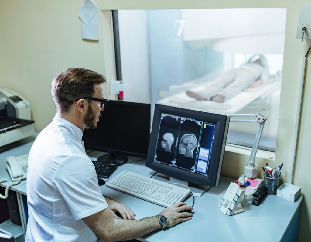
The suspicion of cancer is based on several elements: clinical examination (symptoms), biological examinations (blood tests and analyses), imaging examinations (MRI, radio, scanner, etc.).
Cancer can only be proven by taking a sample of the tumour and analysing it under the microscope by an anatomopathologist.
This study makes it possible to understand the history of cancer cells, the development of tumours and their metastases (secondary focus away from an initial focus) and to classify cancerous tissues according to their nature.

ONCOLOGICAL SURGERY
Surgery is a local cancer treatment that aims to remove the tumour, associated lymph nodes and any metastases. It is also known as the removal of the tumour or cancerous lesion.
Oncology surgeons may perform procedures such as:
- Laparoscopic surgery to remove malignant tumours from the abdomen or pelvis;
- Minimally invasive surgery (laparoscopy) for breast cancer
- Surgery for testicular tumours
- Surgical removal of metastases in internal organs (e.g. lungs).
CHEMOTHERAPY
Chemotherapy (supported in our oncology department) is a treatment that involves the administration of drugs that act on cancer cells, either by destroying them or preventing them from multiplying.
Chemotherapy can be used to treat certain types of leukaemia, lymphoma, breast cancer, lung cancer, head and neck cancer, stomach cancer and Hodgkin’s lymphoma. It can also be used to treat certain non-cancerous (benign) conditions.
Chemotherapy is given intravenously (IV) or as tablets taken by mouth; it is not injected into a vein like antibiotics.
Chemotherapy can be used as the main treatment for cancer, or in addition to other treatments such as radiotherapy and surgery. It can also be used to prevent cancer from coming back after surgery or radiotherapy.

RADIOTHERAPY
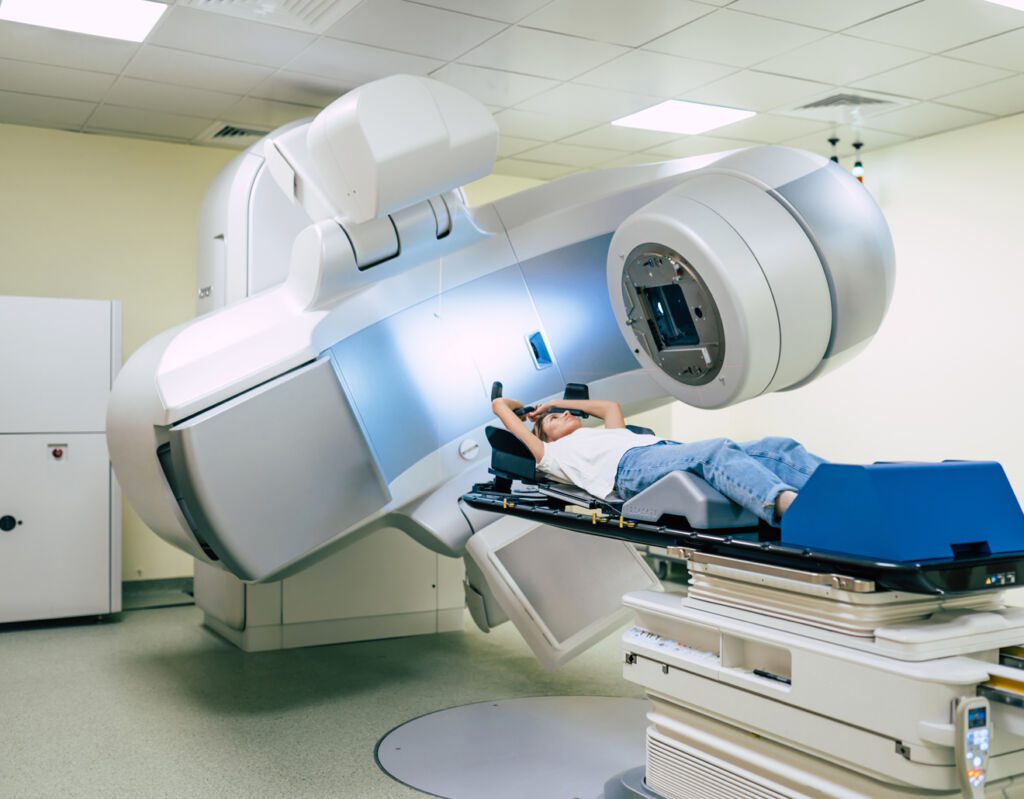
Radiotherapy is a locoregional (specific area or organ) treatment for cancer. It uses radiation to destroy cancer cells by stopping their ability to multiply.
Radiotherapy is used to treat many types of cancer, including certain types of head, neck, brain, lung, breast and skin cancer.
Radiotherapy can be used before surgery to shrink tumours and make them easier to remove. It can also be used after surgery if cancer remains or has spread (metastasised) to other parts of the body.
Radiotherapy damages the DNA of cells so that they cannot divide and develop into new tumours. This can happen in two ways:
- Directly – when the radiation directly affects the site of the tumour
- Indirectly – when the radiation passes through healthy tissue surrounding the tumour.
WHAT IS LYMPHOMA?
Lymphoma (treated in our oncology department) is a type of cancer that develops in the lymphatic system, a network of organs, glands and vessels that helps the body fight infection and other diseases. The lymphatic system includes the spleen, tonsils, adenoids and thymus.
There are different types of lymphoma:
– Hodgkin’s lymphoma: This type of cancer occurs when cells in part of the lymphatic system grow out of control. These cells can spread to other parts of the body through the blood or lymphatic system.
– Non-Hodgkin’s lymphoma (NHL): NHL includes many different types of cancer that are not named according to where they develop in the body. For example, mantle cell lymphoma starts in cells outside the spleen; diffuse large B-cell lymphoma starts in B-cells outside the spleen; and Burkitt’s lymphoma starts in cells in the wall of the stomach called plasma cells.
– Lymphomas are usually treated with chemotherapy or radiotherapy (sometimes both):You may also need surgery or other procedures to remove cancerous tissue from your body before you start treatment or as part of your treatment plan.
WOMB (UTERUS) CANCER
Uterine/ womb cancer (treated in our oncology department) is a disease characterised by the formation of malignant (cancerous) cells in the tissues of the uterus. Uterine cancer is a disease characterised by the formation of malignant (cancerous) cells in the tissues of the uterus.
The most common type of uterine cancer is endometrial cancer, which develops in the lining of the uterus (endometrium). The second most common type of uterine cancer is uterine sarcoma, which develops in the muscle tissue. Cancers that develop outside these normal tissues are much less common than cancers that start in these tissues.
Cancer can also spread from other organs to the uterus, but this type of spread is not called uterine cancer because it is not the cause.
Symptoms include abnormal vaginal bleeding and pelvic pain or pressure that lasts more than two weeks or occurs more frequently than once every two months.
We present to you some articles relating to pathologies treated in our Mediterranean clinic, with an effective and efficient medical and paramedical team.
Adenocarcinoma is a type of cancer that develops in glandular tissue, such as the lining of the lungs, bowel or breast. It can also occur in other organs. Glandular tissue secretes hormones and other substances that control the body’s growth and development.
Adenocarcinoma is a malignant tumour of glandular epithelial cells. The term adenocarcinoma is used to describe a wide variety of cancers that share some characteristics with normal glandular tissue, including a tendency to invade local tissue, spread locally before spreading systemically, and frequently metastasise to lymph nodes or distant sites (such as the lungs or liver).
Symptoms of adenocarcinoma:
Symptoms of adenocarcinoma are generally non-specific and may include:
- Blood in the urine or stool
- Tiredness or weakness
- Weight loss for no apparent reason
- Nausea or vomiting
Skin cancer (treated in our oncology department) is a disease in which malignant cells form in the skin. Melanoma is the deadliest form of skin cancer.
Skin cancer is common, but most cases are curable if caught early. Skin cancer is more likely to develop on areas exposed to the sun, such as the face, ears, lips and neck.
The symptoms of skin cancer vary depending on the type of cancer and where it develops on the body.
The symptoms are as follows:
- A suspicious or developing mole (called a melanoma)
- Wounds that do not heal after a month, especially on the face or hands.
- Other changes in moles that have been there for years.
What causes skin cancer?
Sun’s Ultraviolet (UV) rays from the sun damage the skin’s DNA and cause skin cells to grow abnormally. This damage can lead to skin cancer. Most people are exposed to UV rays every day without realising it because they spend time outdoors, especially during the peak hours of sunlight (10 to 14 hours).
Sun exposure is not limited to outdoor activities such as gardening and golf – even going out for lunch can increase the risk of developing skin cancer. UV exposure time is also important: the more hours you spend outdoors without sunscreen or protective clothing on your arms and legs, the more likely you are to develop skin cancer.
Sarcomas are cancers that start in the body’s connective tissue. Connective tissue is a type of tissue that supports and connects other types of tissue in the body. It is found in organs, muscles, bones, cartilage and blood vessels.
Sarcomas can occur almost anywhere in the body, but they are most commonly found in bones or muscles. They are also called soft tissue sarcomas because they develop in the soft tissues of the body, such as fat or muscle. Cancer cells grow quickly and invade surrounding healthy tissues and organs.
Sarcoma treatment
Sarcomas are rare but aggressive cancers that can affect any part of the body. They are usually treated with surgery followed by chemotherapy or radiotherapy. The type of treatment depends on the origin of the cancer, its severity and whether it has spread to other parts of the body (metastasised).
There are many different types of sarcoma and they are treated differently:
Soft tissue sarcoma (STS) affects fatty and muscular tissues such as tendons and ligaments around joints, bone marrow cells, blood vessels, nerves, fat around internal organs such as the spleen, liver, spinal cord cells and bone marrow cells.
Hodgkin’s lymphoma is a cancer of the lymphatic system, which is part of the body’s immune system. The lymphatic system includes the lymph nodes, spleen and tonsils.
Lymphoma is a cancer that affects the lymphoid tissues that fight infections and other diseases. Lymphoid tissues include the lymph nodes and the spleen. Lymphoma can also occur in other parts of the body, such as the bone marrow and gastrointestinal tract (stomach and intestines).
Hodgkin’s disease is named after the English physician Thomas Hodgkin, who first described it in 1832.
In 1866, the French pathologist Georges de Morsier first described another type of lymphoma called non-Hodgkin’s lymphoma (NHL).
Symptoms of Hodgkin’s lymphoma include:
- Painless swelling of one or more lymph nodes (lymphadenopathy)
- Feeling tired or weak
- Fever
- Difficulty breathing
A brain tumour is a mass of abnormal tissue that develops in the brain. A tumour can be benign or malignant. Most types of tumour can be removed by surgery. Some tumours are inoperable and should be treated with radiotherapy or chemotherapy.
What is a brain tumour?
A brain tumour is an abnormal growth of cells in the brain or spinal cord. The cells grow out of control and form a mass called a tumour.
The term ‘brain tumour’ covers all the different types of lesions that occur in the central nervous system (CNS). These include:
– Benign (non-cancerous) tumours
– Malignant primary lymphomas of the CNS
– Brain tumours can cause a wide range of symptoms depending on their size, location and type of tissue. Some symptoms may include headaches, seizures, vomiting, personality changes, vision problems, and problems with balance and coordination.
Treatment depends on the type and location of the tumour. Surgery is often used to remove the tumour and, if necessary, surrounding tissue. Radiotherapy may also be needed to reduce the risk of recurrence. For some types of brain cancer, chemotherapy may also be recommended after surgery or radiotherapy.
Bone cancer (treated in our oncology department) is a rare disease that occurs when cancer cells form in the bone. The most common types of bone cancer are osteosarcoma, Ewing’s sarcoma and chondrosarcoma.
Bone cancer can occur at any age, but is more common in young adults. Women are more likely to develop the disease than men.
Symptoms of bone cancer include:
– Pain in the bones or joints
– Stiffness or swelling in the joints (especially in the hands and feet)
– A lump or swelling on the skin near a bone (such as the (femur) thigh, arm bone, pelvis or chest) that does not disappear with treatment.
Bone cancer is a type of cancer that develops in the bones. Bone tissue is made up of cells called osteoblasts and osteoclasts that contribute to bone strength and health. Osteoblasts make new bone tissue, while osteoclasts break down old bone tissue and replace it with new bone. If these processes go wrong, they can lead to the development of abnormal cells (tumours) that can invade nearby tissues or spread to other parts of the body (metastases).
There are two main types of bone cancer: primary and secondary:
– Primary tumours develop directly from osteoblasts or osteoclasts in the bone marrow or cortex (outer layer) of long bones, such as those in the arms or legs.
– Secondary tumours can occur when cancer cells from another part of the body enter one of these areas through the bloodstream or lymphatic system and form abnormal tumours.
The Mediterranean clinic is at the forefront of orthopaedic surgery, specialising in the placement of total hip replacements that allow our patients to recover quickly and effectively.
To find out more about oncology and the role of the oncologist, we invite you to read these few lines.
Blood cancer, also known as leukemia, is a serious disease that affects blood cells. Learn about the causes, symptoms and treatment options for blood cancer in this article.
Leukemia is a type of blood cancer that can be difficult to understand. Learn about the causes, symptoms and treatment options in this informative guide.
BIOPSY
A biopsy is a procedure in which a doctor takes a small sample of tissue from your body. The sample is then examined under a microscope to see what type of tumour it is and how far it has spread.
A biopsy may be performed for many reasons, including finding out if you have cancer. If your doctor thinks you have cancer, he or she may recommend a biopsy to confirm the diagnosis.
To find out if an existing tumour has recurred after treatment. Your doctor may want to check your tumour again before deciding on other treatment options.
To check whether certain symptoms – such as swelling or pain – are caused by cancerous growths in the body (such as lymph nodes).
To decide whether radiotherapy is needed after surgery for prostate or breast cancer that has spread (metastasised) to other parts of the body.
EXERESIS
Exeresis is a medical treatment used in oncology. Exeresis is also known as exenteration or radical surgery.
It is a surgical procedure to remove all or part of an organ or tumour. The aim of the surgery is to remove as much of the cancer as possible and prevent it from spreading to other parts of the body.
Excision can be used to treat many types of cancer, including:
– Breast cancer Cervical (neck) cancer
– Oesophageal (lower part of the throat) cancer
– Stomach (stomach) cancer
– Non-small cell lung cancer
THE MYELOGRAM
Myelograms are often used to detect tumours, cysts or other abnormalities of the spinal cord. The procedure involves injecting a contrast agent into the spaces around the spinal cord. The contrast material may be visible on an X-ray, allowing your doctor to see if there are any problems with your spine.
The most common type of myelogram is an X-ray dye study, also known as a myelogram or myelography. During this test, your doctor will inject a liquid containing a dye into one of your veins to fill the spaces around your spinal cord. The dye makes it easier for the doctor to see spinal problems on an X-ray.
Myelography may be performed to check for tumours or other abnormalities in the spine. It may also be performed on patients who are due to have spinal surgery, as it allows doctors to see if there are any problems in this area before surgery.
Pet Scan
This is an imaging test that can be performed to check for cancer or other health problems in your body.
A PET scan (positron emission tomography) is a type of brain scan that can detect certain types of cancer, heart disease and other problems. A small amount of a radioactive tracer is injected into a vein, and a scanner is used to detect the tracer in different parts of the body. The images produced by the tracer show where there are the most blood vessels and how active they are.
The resulting image shows how organs such as the brain and heart are functioning and whether they have an adequate blood supply. It is also used to detect cancers, including those that progress slowly or are difficult to find during surgery or other tests.
In some cases, PET scans can help doctors decide whether a person should be treated with drugs such as chemotherapy or radiotherapy instead of surgery or other procedures.
You ask, our teams answer.
F.A.Q
Oncology is a branch of medicine that deals with the prevention, diagnosis and treatment of cancer.
Oncology is a broad field that encompasses many other disciplines. Scientists and researchers in this field are known as oncologists (or medical oncologists) or cancerologists.
Oncology is not generally disease-specific, but rather the study of malignant tumours (cancer) and their treatment. It focuses on the use of therapeutic agents to destroy malignant cells. This may include surgery, radiotherapy, chemotherapy, immunotherapy or biotherapy.
Cancer is not a single disease, but rather hundreds of diseases classified according to their origin and appearance under the microscope. Cancer can occur anywhere in the body and at any age, but most commonly after the age of 50. About half of all cancers occur in people between the ages of 70 and 79. Cancers are named according to where they are found (such as lung cancer or bowel cancer), how they look under the microscope, or the type of cell which they originate from (such as lymphoma).
Metastasis is the spread of cancer cells from one organ or part of the body to another. Cancer cells can break away from a tumour and travel through the blood or lymphatic system to other parts of the body. Once there, they start to grow and form new tumours. This is called metastasis.
Cancer cells can break away from a tumour and travel through the blood or lymphatic system to other parts of the body. Once there, they start to grow and form new tumours. This is called metastasis.
Cancer cells can break away from a tumour and travel through the blood or lymph system to other parts of the body. Once there, they start to grow and form new tumours. This is called metastasis.
Oesophageal cancer (treated in our Oncology department) is the growth of abnormal cells in the oesophagus, the muscular tube that carries food from the throat to the stomach. The oesophagus lies behind the windpipe (trachea) and the heart and above the stomach.
Oesophageal cancer does not usually cause any signs or symptoms until it has spread to other parts of the body. If you do develop symptoms, they may include indigestion, difficulty swallowing, pain or discomfort in the chest or back, hoarseness, difficulty breathing and weight loss.
Treatment options for oesophageal cancer include surgery, chemotherapy, radiotherapy and targeted therapy. Surgery is used to remove all or part of the oesophagus containing cancer cells. It can be performed by open surgery (with an incision) or endoscopy (using an instrument called an endoscope). Chemotherapy uses drugs to kill cancer cells by stopping them from dividing properly or by damaging their DNA. Radiotherapy uses high energy beams directed at cancerous tissue to kill cancer cells and shrink tumours. Targeted therapy uses drugs that target proteins produced by certain types of cancer cells so that they can be destroyed by your immune system.
Laparoscopic surgery, also known as minimally invasive surgery, is a type of surgery in which fine tools and instruments are inserted through small incisions (cuts) in the body. The surgeon uses these tools to see and reach inside the body.
Laparoscopy is usually performed through several small incisions in the abdominal wall. A laparoscope is a thin tube with a camera at one end and a light source at the other. It allows the surgeon to see inside the abdomen or pelvis during the operation.
Laparoscopic surgery can be used to treat many conditions of the abdomen, pelvis or reproductive organs, such as:
- Appendicitis
- Gallstones
- Stomach ulcers
- Kidney stones
- Bowel obstructions (blockages)
- Uterine fibroids
Cancer is a disease that affects people of all ages. The most common cancers are breast cancer, prostate cancer, lung cancer, colorectal cancer, melanoma and non-melanoma skin cancer, as well as kidney and renal pelvis cancer (treated in our Oncology department).
The most commonly used biological tests for cancer diagnosis:
– Tumour marker testing:
Tumour markers are substances that can be found in the blood or urine. They can be associated with certain types of cancer and can help your doctor determine whether you have cancer, how far it has spread and what type it is. Screening for tumour markers is often performed with a simple blood test.
– Looking for specific mutations:
Some cancers have specific mutations that can be identified through testing. These mutations can help your doctor decide what type of treatment you need.
Biomarkers are proteins or molecules that can be found in a person’s blood, urine, saliva or other body fluids. They can be found in small amounts and often provide information about the presence of a disease or a person’s risk of developing a disease. Biomarkers are often used in the diagnosis of cancer.
The most common type of biomarker is a protein that is produced by cancer cells and circulates in the blood (serum). This protein can be released by damaged cells (such as liver cells), but is not normally present in the blood. Biomarkers for other types of cancer include DNA mutations in blood samples and genetic changes in cells found in stool samples.
Detecting these biomarkers is called biomarker testing or molecular profiling. The aim is to identify specific changes in genes, proteins or other molecules caused by cancer or other diseases, so that patients can receive appropriate treatment according to their individual risks and needs.
This is the swelling of tissues, usually due to the accumulation of fluid. Swelling is a common symptom of many conditions, including infections, inflammation and neoplasms (cancerous tumours).
The cause of swelling is usually easy to identify. In bacterial infections, for example, the surrounding area becomes inflamed as white blood cells attack the bacteria and multiply in the blood vessels, which become clogged with inflammatory cells and proteins. This causes swelling, pain and redness. In some types of cancer, such as lymphoma, the lymph nodes become enlarged because lymphocytes (a type of white blood cell) build up. This can cause swelling in other parts of the body because the lymph vessels become blocked.
Swelling can also occur if there is an abnormal increase in fluid or protein in an organ or tissue. For example, in some types of heart failure, excess fluid accumulates in the lungs or abdomen because the heart cannot pump enough blood to these areas.


